



Echogenic Nerve Block Needles: How 360-Degree Laser-Engraved Micro-Structures Improve Ultrasound Visibility
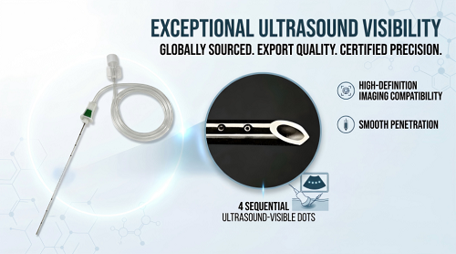
Ultrasound-guided nerve numbing requires clear needle tip views during deep block procedures. Standard smooth metal surfaces often lose sound signals at steep angles. Advanced nerve block needles solve this problem using 360-degree laser-engraved micro-structures. These precise laser-etched micro-geometry arrays act as light-returning sound reflectors. They bounce ultrasound energy directly back to the probe face, improving ultrasound imaging.
This full-circle echogenic needle design gets rid of blind spots during rotation. Doctors gain better ultrasound visibility, constant viewing, and reliable placement tracking. The bright echogenic needle offers high contrast against surrounding soft tissues. Medical staff easily spot delicate tissue structures, hit exact nerve targets, and finish ultrasound-guided procedures with high confidence.
Key Takeaways
Laser-engraved tiny patterns reflect sound waves back to the probe, making the needle very easy to see.
A complete 360-degree design eliminates hidden dark areas when the needle spins within the body's tissue.
Strong medical-grade 304 stainless steel helps the needle stay tough yet bendable during deep procedures.
Clear ultrasound pictures help doctors stay away from blood vessels and safeguard delicate target nerves.
Physics of Acoustic Reflection in Regional Anesthesia
Specular Reflection vs Echogenic Needle Scattering
High-frequency ultrasound waves travel through soft tissue layers during clinical nerve block delivery. Smooth metallic surfaces act like flat optical mirrors for acoustic energy. Standard smooth steel cannulas reflect sound waves away from the receiver probe. Steep insertion angles increase acoustic signal loss dramatically during deep tissue navigation. Entry angles beyond 30 degrees cause severe sound wave scatter away from the transducer face. Returning acoustic signals fail to hit the ultrasound array sensors. Anesthesiologists lose track of the smooth metal cannula during steep advancement. Clinicians lose sight of the target nerve location and surrounding tissue structures. Poor shaft visibility increases procedural difficulty during deep target approaches. Attenuation limits clear needle imaging in complex clinical scenarios.
Unmodified metallic surfaces create extreme physical acoustic impedance boundaries against soft biological tissue. Sound waves travel quickly through steel but move slowly through human tissue. This sharp density interface reflects sound waves away at predictable geometric angles. Smooth metal shafts scatter incident sound energy away from the probe face when inserted obliquely. Consequently, conventional instruments disappear on the monitor screen during steep peripheral injections. An echogenic needle eliminates signal loss by scattering acoustic energy back to the ultrasound transducer array sensors.
Clinicians evaluate signal attenuation by measuring acoustic echo intensity across variable insertion angles. Smooth metallic surfaces suffer from severe specular reflection at oblique approach angles. An echogenic needle counters severe acoustic signal attenuation during regional anesthesia interventions by scattering sound energy effectively.
Measurement Parameter | Smooth Surface Mechanism | Echogenic Surface Mechanism |
|---|---|---|
Surface Reflection Behavior | Steep insertion angles reduce visibility through strong specular reflections away from the ultrasound probe. | Micro-textures scatter wave energy back along incident pathways to maintain continuous visibility. |
Echo Contrast Data | An ex vivo study shows a Contrast-to-Noise Ratio drop from ~30 dB at 0° to ~9 dB at 60° insertion angle. | Precision micro-geometry preserves high signal intensity and echo return at steep insertion angles. |
Micro-Geometric Retroreflection Dynamics
Engineers modify outer cannula boundaries to overcome physical specular reflection limits. Micro-textured surface modifications create intentional acoustic impedance mismatches along the metallic shaft. These etched micro-geometric surface arrays function as microscopic corner-cube retroreflectors. The precise surface geometry captures incoming sound waves directly. It redirects wave energy back along the exact incident pathway regardless of insertion angle. This physical reflection mechanism guarantees reliable visualization during ultrasound-guided regional anesthesia. Using an echogenic needle guarantees accurate tip localization.
A specialized echogenic needle uses retroreflective dynamics to return sound waves to the receiver array. Acoustic waves bounce directly back to the active transducer face. This physical reflection mechanism enhances overall ultrasound imaging quality within dense tissue layers. Target nerve fibers stand out clearly against adjacent anatomical structures during regional anesthesia procedures. Clinicians monitor the moving shaft during real-time needle adjustments. Accurate placement protects delicate nerve tissue from accidental mechanical trauma. Consistent acoustic backscatter provides stable imaging contrast across broad trajectory angles.
Furthermore, reliable imaging performance depends on specialized surface patterns. An echogenic needle preserves acoustic wave coherence across steep insertion paths. Controlled sound scattering optimizes overall ultrasound display brightness. Anesthesiologists receive clear imaging feedback while maneuvering near critical target nerve locations. Continuous acoustic wave return maintains clear needle visibility throughout regional anesthesia blocks. The echogenic needle performance simplifies complex point-of-care needle placement tasks for medical staff.
360-Degree Engraving for Enhanced Ultrasound Visibility
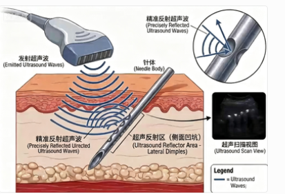
Eliminating Rotational Dead Zones for Constant Visibility
Standard puncture tools lose clear visibility during rotation under ultrasound guidance. Clinicians often rotate the echogenic needle shaft during complex deep block procedures. Traditional designs feature uneven surface etching along the metal shaft. These uneven patterns create dark rotational dead zones on the ultrasound monitor screen. As a result, anesthesiologists experience intermittent signal loss during critical nerve block procedures. Advanced surface technologies solve this spatial tracking limitation completely. Engineers place specialized surface structures along the entire shaft circumference to provide uniform acoustic backscatter.
The optimized needle design features a continuous 360-degree laser-engraved pattern. Manufacturing teams engrave precision 4x4 arrays of micro-structures along the distal shaft and tip. This full 360-degree distribution ensures continuous wave scattering regardless of axial rotation. Sound waves bounce back directly to the ultrasound transducer face at every trajectory angle. Anesthesiologists maintain instant real-time needle visibility during complex tissue maneuvers. The constant signal return guarantees exceptional needle tip visualization near sensitive target nerve bundles. Clinicians achieve enhanced ultrasound visibility, improving overall procedure speed during regional anesthesia interventions. Reliable imaging performance depends on continuous acoustic wave feedback across all rotation angles. High acoustic reflection guarantees stable visibility throughout the peripheral nerve block procedure. Ultrasound machines display high-contrast signals as the shaft approaches target anatomical layers. Continuous wave scattering eliminates target loss during delicate angle adjustments.
Precision Micro-Arrays and Structural Integrity
An echogenic needle maintains high shaft visibility during difficult deep approaches. Clinicians flex the strong steel shaft during targeted nerve block adjustments. High elasticity prevents permanent shaft bending or catastrophic instrument failure in deep tissue layers. Manufacturers craft the echogenic needle shaft using 304 medical-grade stainless steel. Highly precise laser beams create detailed micro-grooves without compromising overall metal strength. The focused thermal energy modifies only a thin surface layer along the outer lumen to protect structural integrity.
Key Aspect | How It Preserves Mechanical Properties |
|---|---|
Localized Energy | The laser beam focuses energy only on the mark area, minimizing thermal impact on the component. |
Annealing Marking Technique | Controlled heating forms an oxide layer on the surface without significant material removal, keeping the surface intact. |
Contrast with Material Removal | Annealing marking on austenitic 304 steel modifies only a thin surface layer, preserving the bulk material structure. |
General Outcome | The process maintains functional characteristics, including high elasticity and overall shaft durability. |
Precision micro-etching provides clear real-time ultrasound imaging without weakening the cannula shaft. An echogenic needle maintains structural durability during demanding regional anesthesia interventions. High acoustic contrast optimizes overall needle imaging during ultrasound guidance during deep tissue navigation. Clinicians track the moving shaft across dense anatomical structures with exceptional visual feedback. Clear visualization prevents accidental trauma to surrounding nerve tissues. Reliable echogenic needle performance delivers enhanced needle imaging during critical point-of-care administration. Enhanced ultrasound visibility allows medical staff to execute precise injections near delicate nerve bundles. Continuous acoustic wave reflection ensures stable visibility across variable insertion approach angles. Superior imaging clarity guides clinicians safely toward target nerve locations during every insertion. An echogenic needle combines mechanical elasticity with bright visual clarity.
Clinical Safety in Ultrasound-Guided Procedures
First-Pass Success in Peripheral Nerve Block Needles
Echogenic technology offers clear medical advantages throughout regional anesthesia treatments. Ultrasound imaging allows doctors to track the moving shaft easily inside deep tissue. Bright contrast helps healthcare teams guide the needle directly toward target nerve pathways. An echogenic needle scatters sound waves straight to the sensor probe. This scattering improves general visibility while performing complicated nerve blocks.
Medical trials check first-try success rates using different cannula models. Research on vein access showed a 69% success rate on the first attempt with an echogenic needle. Standard tools reached an 80% success rate in the same study with a p-value of 0.22. These numbers prove there is no real mathematical difference between the two tools. Still, clear needle tip visibility always helps clinicians hit exact nerve targets safely. Dependable visual feedback boosts overall accuracy when making careful adjustments.
Mitigating Vascular Traumatization and LAST Risks
Keeping track of the needle shaft protects patients during deep nerve block procedures. Doctors need to identify tissue layers accurately while using ultrasound guidance. Poor visibility raises the danger of accidentally hitting deep blood vessels. Clear ultrasound images help workers navigate safely away from delicate blood channels. Live tracking lowers the chance of injecting medicine into blood vessels by mistake. Avoiding direct blood stream entry prevents dangerous toxic reactions to local painkillers.
Special tip shapes give doctors better physical control while giving nerve blocks. A dual back-bevel tip design gently moves tissue fibers apart during entry. Smooth points lower pushing force and reduce physical tissue damage. Medical pain studies prove multi-bevel tips cut patient pain scores by 36 mm on the visual scale. Lower pushing force keeps touch sensitivity sharp for exact nerve targeting. Doctors feel tissue layer changes clearly as the shaft moves forward. Great echogenic needle performance provides high accuracy, lifting safety levels for ultrasound-guided treatments.
Comparing Echogenic Technologies for Regional Anesthesia
Limitations of Dimpled and Coated Needles
Doctors need clear pictures when using sound waves to numb nerves. Special coatings and metal dents try to bounce sound back better. However, plastic coatings can peel off inside deep tissue layers. Metal dents bounce sound waves in uneven directions. These older tools do not show the needle clearly all the time. Doctors lose sight of the needle at steep entry angles.
An echogenic needle fixes these problems using laser-etched micro-patterns. Precise laser tools cut tiny geometric shapes into the metal shaft. These micro-patterns bounce sound waves straight back to the machine. An echogenic needle stays bright and clear at any angle. Good images help doctors guide the needle close to target nerves.
Tactile Control and Workflow Integration
A well-designed needle helps doctors work faster during nerve numbings. An echogenic needle reflects sound waves clearly without changing how the metal feels.
Target Tracking: Laser-etched microstructures reflect ultrasound waves, improving needle tip visualization and safety.
Streamlined Workflow: Clear visualization reduces unnecessary repositioning steps, allowing a smooth approach toward the target nerve.
Target Placement: Accurate sound wave reflection ensures precise needle placement next to critical nerve bundles.
Doctors gain better control by combining bright sound wave pictures with smart engineering.
Design Feature | Purpose | Relation to Tactile Feedback |
|---|---|---|
360° X-pattern Laser Engraving | Improves ultrasound visibility and tip placement. | It does not affect physical feel; its role focuses purely on visual enhancement. |
30° Back-Cut Bevel Design | Provides consistent puncture feel. | This specific bevel geometry preserves the familiar tactile feedback for nerve tissue. |
Combined Design Principle | Separates visual and tactile functions. | Laser structures handle acoustic reflection, allowing the cannula bevel to maintain tactile feedback near the nerve during regional anesthesia. |
A smart needle design helps doctors find target nerves easily. An echogenic needle stays bright during deep nerve numbing steps. Medical teams place nerve block needles with great speed and care. Clear images give workers full confidence during sound-guided nerve care.
Exact 360-degree laser-etched micro-patterns send ultrasound signals straight back to sensors. This special process fixes weak sound waves during nerve block steps. Custom nerve block tools remove dark hidden spots entirely. An echogenic needle keeps a clear picture even at sharp entry angles. Doctors watch the moving tip close to target nerve fibers without dropping video feedback.
Easy shaft viewing speeds up nerve tracking and protects patients during regional block procedures. Flexible 304 stainless steel strengthens the whole tool during direction changes. A gentle tip shape pushes tender body parts aside safely. This echogenic needle pairs clear ultrasound pictures with medical protection for local numbing shots. Great needle visibility sets a strong goal for modern ultrasound nerve care.
FAQ
How do micro-structures improve needle ultrasound visibility?
Laser-carved tiny patterns bounce sound signals right back toward the sensor. Sending these waves back makes the metal shaft far easier to see on-screen during numbing treatments. Spotting the tip clearly helps medical staff follow the path of the tool as it moves through body tissue, keeping the point in view so it stays on the right course.
Why does a 360-degree needle design improve nerve block precision?
Covering the whole shaft with a full-circle pattern gets rid of dark blind spots when the tool turns. Doctors can twist the instrument any way they need without blurring the live picture on the monitor. Because the shaft stays bright from every angle, workers can safely guide the point past delicate nerve fibers to put it in the exact right spot.
How does needle tip geometry enhance safety during ultrasound-guided procedures?
A specially angled tip smoothly parts tissue layers instead of tearing them. Shaping the point this way lowers pushing effort while giving the doctor better physical control over the tool. Medical teams can easily steer close to target nerves without causing accidental harm during careful image-guided procedures.
What material constructs a flexible echogenic needle?
Makers craft this clear-view tool out of medical-grade 304 stainless steel. Using this tough metal gives the shaft great springiness when navigating tricky paths through the body. This sturdy build stops the instrument from snapping, keeps its shape strong, and holds it steady under live ultrasound imaging.
Please Contact Us!
Recent News
1.What Are the Different Types of Nerve Block Needles?
2026-08-052.Ultrasound Guided Nerve Block Needle: Design, Benefits, and Clinical Applications
2026-08-043.What Is an Echogenic Needle for Ultrasound Guided Nerve Blocks?
2026-07-204.What Needle Is Used in Regional Anesthesia?
2026-07-155.Ultrasound Visible Needle vs Standard Needle: What Is the Difference in Ultrasound-Guided Procedures?
2026-07-156.Why Choose ShineArk as Your Ultrasound Nerve Block Needle Supplier?
2026-07-097.What Size Needle for Femoral Nerve Block?
2026-07-018.What Is the Use of Puncture Needles?
2026-06-299.What Kind of Needle Is Used for a Peripheral Nerve Block?
2026-06-29