



Ultrasound Guided Nerve Block Needle: Design, Benefits, and Clinical Applications
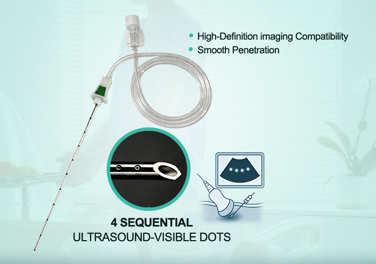
A nerve block needle is a specialized medical device designed to deliver local anesthetic close to targeted nerves during regional anesthesia procedures. Unlike the needle itself blocking nerve signals, the main therapeutic effect comes from the anesthetic medication spreading around the nerve to temporarily interrupt pain transmission.
With the development of ultrasound-guided regional anesthesia, clinicians can visualize anatomical structures in real time, including nerves, blood vessels, fascia, and the advancing needle tip. This improves the accuracy of needle placement and helps reduce potential complications during peripheral nerve block procedures.
Modern ultrasound guided nerve block needles combine precise needle design with enhanced visualization technology. Features such as echogenic surfaces, optimized bevel structures, and high-quality stainless steel construction help clinicians identify needle position more effectively under ultrasound imaging.
Guidance Approach | Clinical Advantage | Key Benefit |
|---|---|---|
Ultrasound Guidance | Real-time visualization of nerves, vessels, and needle trajectory | Supports more accurate needle placement compared with landmark-based techniques |
Traditional Landmark Technique | Relies mainly on external anatomical references | May provide less information about individual anatomical variations |
Clinical outcomes can vary depending on the block type, patient anatomy, equipment quality, and operator experience. However, ultrasound guidance has become an important technique for improving visualization and supporting safer regional anesthesia practice.
Key Takeaways
Nerve block needles deliver local anesthetic near targeted nerves rather than directly blocking nerves themselves.
Ultrasound guidance provides real-time visualization of nerves, vessels, and needle movement.
Echogenic needles improve needle visibility by enhancing ultrasound reflection.
Proper needle tip positioning near the target nerve helps achieve effective anesthetic distribution.
In-plane ultrasound techniques allow continuous visualization of the needle shaft and tip.
High-quality nerve block needles typically feature medical-grade stainless steel, optimized bevel designs, and sterile single-use construction.
Understanding Nerve Block Needles
A nerve block needle is engineered to accurately deliver local anesthetic around a specific nerve or nerve plexus. During a peripheral nerve block procedure, the clinician advances the needle toward the target anatomical location while using ultrasound imaging or other guidance methods to confirm positioning.
The goal is not to penetrate the nerve, but to position the needle tip adjacent to the nerve so the anesthetic solution can spread around the intended area. This approach supports effective pain management while reducing unnecessary tissue disturbance.
Structural Anatomy and Materials
The design and material quality of a nerve block needle directly influence handling performance during regional anesthesia procedures. Most high-quality needles are manufactured using medical-grade stainless steel, such as 304 stainless steel, which provides a balance of strength, flexibility, and corrosion resistance.
Material Property | Performance Characteristics | Clinical Importance |
|---|---|---|
Medical Stainless Steel | High strength and corrosion resistance | Provides reliable needle integrity during insertion |
Flexible Needle Shaft | Balanced rigidity and flexibility | Supports controlled advancement through different tissue layers |
In addition to the needle shaft, features such as the hub design, extension tubing, and connection system influence procedural convenience. A stable connection between the needle and injection system helps minimize unnecessary needle movement during anesthetic administration.
Non-Cutting Bevel Configurations
Needle bevel design plays an important role in tissue interaction. Many regional anesthesia needles use specialized bevel geometries designed to support controlled advancement and minimize unnecessary tissue disruption.
Clinical Tip: The needle tip should be carefully visualized and controlled throughout advancement to maintain appropriate positioning near the target nerve.
Selecting the appropriate bevel design depends on the clinical application, target depth, and clinician preference. Proper ultrasound visualization remains essential regardless of needle tip configuration.
Echogenic Micro-Surface Engineering
Ultrasound visibility is one of the most important considerations when selecting a modern nerve block needle. During ultrasound-guided regional anesthesia, the clinician needs to identify the needle shaft and tip clearly while advancing toward the target nerve.
Conventional smooth needle surfaces may produce weaker ultrasound reflections, especially when the insertion angle is steep. Echogenic nerve block needles are designed with specialized surface structures that improve acoustic reflection and make needle visualization easier during procedures.
ShineArk AN-N/AN-M Series echogenic needles use laser-etched microstructures to enhance ultrasound signal reflection. These micro-patterns are designed to improve tip identification and support accurate needle tracking under real-time imaging.
The laser-etched surface technology provides several advantages:
Enhanced Ultrasound Reflection: Micro-structured surfaces increase acoustic signal return, improving needle visibility on ultrasound monitors.
Improved Needle Tip Identification: Clearer visualization helps clinicians confirm needle position relative to the target nerve.
Better Visualization at Different Angles: Optimized surface design supports improved visibility during challenging needle approaches.
By combining advanced needle geometry with ultrasound imaging, clinicians can better monitor needle advancement and maintain appropriate distance from sensitive anatomical structures.
Key ultrasound performance features include:
Enhanced Sonographic Visibility: Built with advanced echogenic technology to improve needle visualization.
Micro-Reflective Surface Structure: Laser-etched patterns improve ultrasound signal reflection.
Reliable Tip Recognition: Helps clinicians distinguish the needle tip from surrounding tissues during ultrasound-guided procedures.
Benefits of Ultrasound Guidance in Regional Anesthesia
Ultrasound guidance has changed the workflow of regional anesthesia by allowing clinicians to visualize anatomical structures in real time. Compared with traditional landmark-based techniques, ultrasound provides additional information about nerves, blood vessels, fascia, and surrounding tissues.
The use of ultrasound guidance helps clinicians make informed decisions about needle trajectory, target location, and anesthetic distribution.
Real-Time Anatomical Visualization
Individual anatomy can vary significantly between patients. Ultrasound imaging allows clinicians to identify important structures before and during needle advancement.
Real-time visualization helps with:
Identifying the target nerve or nerve plexus.
Recognizing nearby blood vessels and avoiding unnecessary contact.
Observing the spread of local anesthetic around the target area.
Clinical Factor | Impact of Ultrasound Guidance |
|---|---|
Anatomical Variation | Allows clinicians to identify patient-specific structures before needle placement. |
Needle Position Control | Provides real-time feedback during needle advancement. |
Injection Monitoring | Helps observe anesthetic spread around the target area. |
Tracking Needle Trajectory and Tip Position
Accurate visualization of the needle trajectory is essential for ultrasound-guided nerve blocks. The clinician must continuously monitor the relationship between the needle tip and surrounding anatomical structures.
Several factors can influence needle visibility:
Out-of-Plane Approach: The needle may appear as a small point instead of a continuous shaft image, making tip identification more challenging.
Insertion Angle: Extremely steep angles may reduce ultrasound reflection from conventional needle surfaces.
Needle Diameter: Smaller needles may produce weaker ultrasound reflections.
Probe Alignment: Proper ultrasound probe positioning helps optimize image quality.
Techniques that improve needle visualization include:
In-Plane Needle Approach: Keeps the needle aligned with the ultrasound beam and allows visualization of the needle shaft and tip.
Probe Adjustment: Tilting or repositioning the probe can improve ultrasound beam alignment.
Echogenic Needle Technology: Specialized surface structures improve acoustic reflection and needle visibility.
Modern echogenic needles support better visualization during difficult procedures by improving the contrast between the needle and surrounding tissues.
Monitoring Local Anesthetic Distribution
Ultrasound guidance allows clinicians to observe local anesthetic spread during injection. This helps confirm whether the medication is moving toward the intended anatomical area.
Clinical Practice Note: Clinicians should confirm appropriate anesthetic distribution and continuously monitor patient response during nerve block procedures.
Real-time imaging provides additional procedural information by showing tissue movement and fluid distribution. If the anesthetic does not spread as expected, the clinician can adjust needle position or injection strategy.
This approach supports controlled medication delivery while maintaining attention to patient safety and procedural accuracy.
Enhancing Safety in Regional Anesthesia
Ultrasound-guided regional anesthesia improves procedural control by providing direct visualization of anatomical structures. Instead of relying only on surface landmarks, clinicians can observe nerves, vessels, fascia, and needle movement during the procedure.
Using ultrasound guidance helps support safer needle advancement and more controlled local anesthetic delivery.
Preventing Unintended Nerve or Vascular Contact
During peripheral nerve block procedures, accurate needle positioning is important. Ultrasound visualization allows clinicians to monitor the relationship between the needle tip, target nerve, and nearby vascular structures.
Track needle advancement toward the target anatomical area.
Identify surrounding structures before injection.
Observe anesthetic spread during administration.
Important Safety Note: Ultrasound guidance supports visualization, but successful regional anesthesia also depends on proper training, clinical judgment, and standardized safety practices.
Optimizing Local Anesthetic Delivery
Ultrasound guidance allows clinicians to visualize where anesthetic is deposited around the target nerve. This may help optimize medication volume according to the specific procedure, patient condition, and clinical requirements.
Appropriate anesthetic management requires consideration of multiple factors, including block location, drug selection, patient characteristics, and practitioner experience.
Improving Procedural Efficiency
Clear ultrasound images can reduce unnecessary needle repositioning and help clinicians maintain a controlled approach during regional anesthesia procedures.
However, clinical outcomes depend on many factors, including the type of nerve block, patient anatomy, ultrasound equipment, and operator expertise.
Types of Nerve Block Needles
Different types of nerve block needles are designed for specific regional anesthesia applications. Selection depends on the procedure, target depth, ultrasound technique, and clinician preference.
Needle Type | Main Characteristics | Common Applications |
|---|---|---|
Echogenic Nerve Block Needle | Features ultrasound-enhanced surface structures for improved needle visibility. | Ultrasound-guided peripheral nerve blocks. |
Stimulating Nerve Block Needle | Designed for procedures using nerve stimulation techniques. | Regional anesthesia procedures requiring electrical nerve localization. |
Standard Regional Anesthesia Needle | Conventional needle design for selected anesthesia applications. | Various injection and regional anesthesia procedures. |
Key Features of Ultrasound Guided Nerve Block Needles
When selecting an ultrasound guided nerve block needle, healthcare providers and distributors typically consider needle visibility, material quality, size options, and manufacturing reliability.
Echogenic Needle Surface: Specialized surface structures improve ultrasound reflection and support clearer needle visualization.
Medical-Grade Stainless Steel: High-quality stainless steel provides strength, flexibility, and corrosion resistance.
Multiple Gauge and Length Options: Different needle sizes support various block locations and anatomical depths.
Optimized Bevel Design: Needle tip geometry supports controlled advancement through tissue layers.
Sterile Single-Use Design: Disposable construction supports infection control requirements in clinical environments.
Why Choose ShineArk Nerve Block Needles?
ShineArk develops ultrasound-guided needle solutions designed for regional anesthesia applications. The product range focuses on improving needle visibility, procedural control, and consistent manufacturing quality.
Key product advantages include:
Laser-etched echogenic surface technology for improved ultrasound visibility.
Medical-grade stainless steel construction for reliable mechanical performance.
Support for different needle specifications and OEM customization requirements.
Manufacturing experience focused on ultrasound-guided medical needle products.
FAQ
What is a nerve block needle used for?
A nerve block needle is used to deliver local anesthetic near targeted nerves during regional anesthesia procedures. The needle helps clinicians accurately position medication around the nerve to provide temporary pain relief.
Does a nerve block needle enter the nerve?
No. During standard peripheral nerve block procedures, the needle tip is generally positioned adjacent to the target nerve rather than intentionally inserted into the nerve. Ultrasound guidance helps clinicians monitor needle location and surrounding anatomy.
Why are echogenic nerve block needles used in ultrasound-guided procedures?
Echogenic nerve block needles use specially designed surface structures that improve ultrasound reflection. This makes the needle shaft and tip easier to identify during ultrasound-guided regional anesthesia.
Why is ultrasound guidance preferred for nerve block procedures?
Ultrasound guidance provides real-time visualization of nerves, vessels, tissues, and needle movement. This additional information helps clinicians perform procedures with better anatomical awareness compared with techniques based only on surface landmarks.
What needle sizes are available for peripheral nerve blocks?
Nerve block needles are available in different gauges and lengths depending on the target nerve location and procedure requirements. Common options include different needle diameters and lengths for superficial and deep regional anesthesia applications.
What kind of needle is used for a peripheral nerve block?
Peripheral nerve blocks commonly use specialized regional anesthesia needles designed for controlled advancement and accurate anesthetic delivery. For ultrasound-guided procedures, echogenic needle designs are often selected to improve visualization.
Please Contact Us!
Recent News
1.Ultimate Guide to Ultrasound-Guided Nerve Block Needles: Safe, Precise, and Disposable Solutions
2026-05-292.What's a Nerve Block Needle, and How Does It Work?
2026-06-083.How to Select the Right Peripheral Nerve Block Needle
2026-06-124.Why Choose ShineArk as Your Ultrasound Nerve Block Needle Supplier?
2026-07-095.What Size Needle for Femoral Nerve Block?
2026-07-016.What Is the Use of Puncture Needles?
2026-06-297.What Is an Echogenic Needle for Ultrasound Guided Nerve Blocks?
2026-07-208.What Needle Is Used in Regional Anesthesia?
2026-07-15