



What Is an Ultrasound Needle for Regional Anesthesia and Biopsy
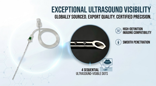
An echogenic ultrasound needle has a tiny textured surface that makes it easy to see on a live ultrasound screen. Normal needles do not have these small reflective patterns. Doctors carefully guide this special tool near sensitive nerves when numbing a specific area of the body. They can also safely collect tissue samples during a focused biopsy, which improves patient comfort and makes the procedure more accurate.
Key Takeaways
Special designs on the surface help doctors see the ultrasound needle clearly on a live screen.
Doctors safely steer the needle to numb nerves or gather tissue samples while avoiding nearby blood vessels.
Seeing the needle inside the body in real time lowers patient pain and helps them heal much faster.
What Is an Ultrasound Needle and How Does It Work?
Micro-Structure Design and Acoustic Reflection
Specialized manufacturing transforms a standard metal shaft into an echogenic ultrasound needle. Engineers engrave microscopic reflective surface patterns onto the outer steel surface.
The laser-engraved micro-structures function as microscopic corner-cube retroreflectors that capture incoming ultrasound waves and redirect them back along the exact incident pathway, regardless of insertion angle. The continuous 360-degree pattern etched along the distal shaft and tip ensures wave scattering at every axial rotation, eliminating blind spots and guaranteeing reliable needle tip visualization at any trajectory angle.
ShineArk utilizes a 4x4 array micro-structure on its AN-N/AN-M Series to maintain this acoustic signal.
High-elasticity 304 medical-grade stainless steel provides essential structural integrity. Moderate strength maintains sharpness and rigidity during target tissue entry. Strong corrosion resistance lowers shaft breakage risks during complex interventions.
Patient safety requires strict international quality standards:
ISO 13485 certification for quality management systems
Sterilization processes complying with ISO 11135 and ISO 11607 for packaging
MDR traceability through UDI tracking from manufacturer to patient
Real-Time Visualization Under Ultrasound Imaging
Real-time guidance lets clinicians track the needle tip through soft tissue layers. High-frequency sound waves bounce off the micro-structures, displaying bright highlights on the monitor. Clinicians view nearby blood vessels and nerve bundles simultaneously to prevent accidental vascular puncture.
Visual guidance elevates procedure success rates. In a phantom model simulating regional anesthesia, passive guidance technology improved first-pass completion from 76.4% (214/280) to 88.9% (249/280), a significant improvement of 16.4% (P = 0.001). Clear tracking under continuous ultrasound imaging shortens procedure time and protects sensitive patient structures.
Clinical Applications: Regional Anesthesia and Ultrasound Guided Needle Biopsy
ShineArk Nerve Block Needle for Regional Anesthesia
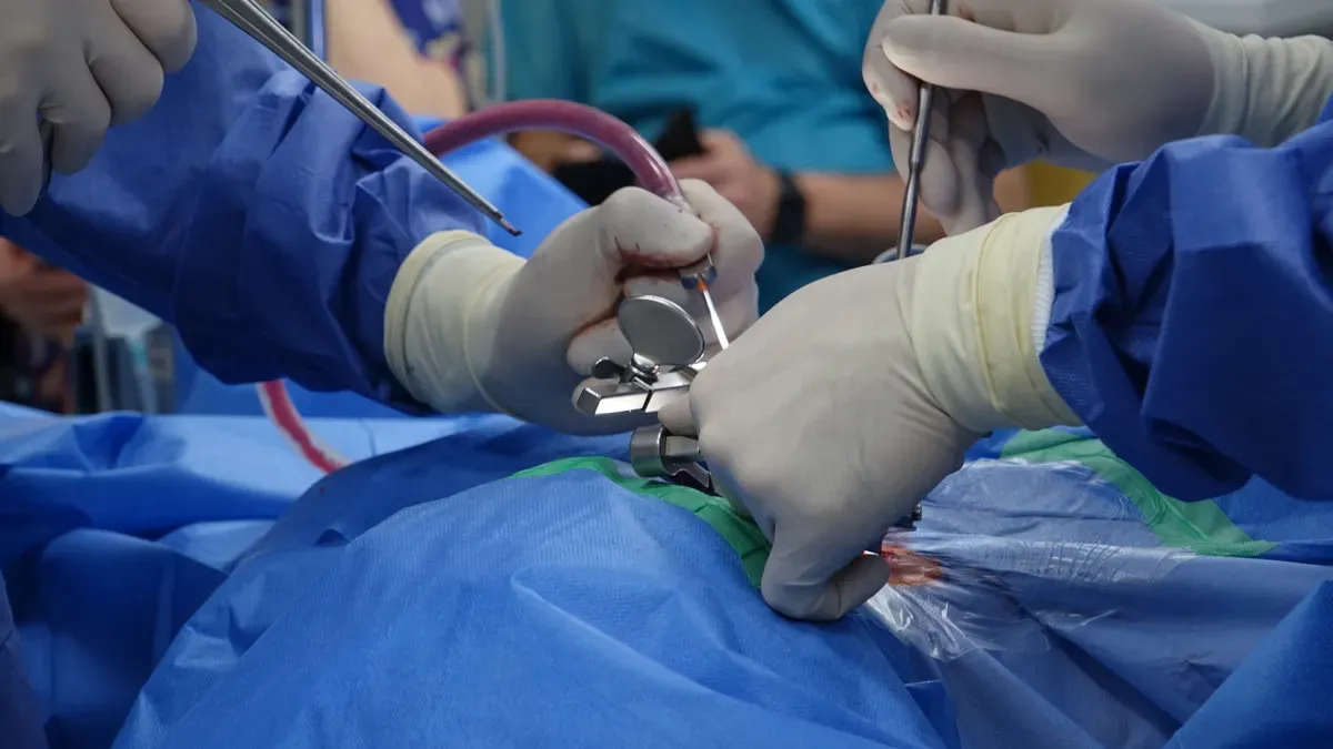
Doctors use the ShineArk AN-N/AN-M Series specialized device for nerve blocks and regional pain control. A double-slanted tip design carefully moves soft tissue fibers aside instead of slicing them. This safe method guards delicate patient nerves effectively while cutting down on physical harm as the needle goes in.
The attached 500mm TPU extension tube lets doctors inject numbing medicine cleanly and safely. Smooth fluid flow prevents sudden pressure jumps during the injection process. As a result, steady medicine delivery speeds up numbing action while greatly reducing dangerous side effect risks.
Performing Ultrasound Guided Needle Biopsies with Precision
Specialists often do an ultrasound guided needle biopsy to check hidden target spots closely. Live ultrasound pictures offer clear moment-by-moment tracking of an echogenic ultrasound needle inside body tissue. Doctors easily pinpoint deep tissue problems without hurting nearby blood vessels under direct ultrasound guidance.
Simple tissue testing relies deeply on accurate placement when taking samples out. Doctors perform fine needle aspiration to draw liquid and cell samples from a shallow spot. Meanwhile, a core needle biopsy cuts out solid tissue pieces from suspicious inner organ lumps for testing.
Clear ultrasound pictures show soft body parts plainly, helping the needle move smoothly through complex areas. Live tracking boosts diagnostic success rates while cutting overall procedure times across treatments. Constant imaging ensures exact placement right inside the target biopsy spot. Finally, modern imaging gives dependable health answers during every careful biopsy test.
Patient Benefits and Procedural Safety Advances
Enhanced Target Accuracy and Complication Reduction
Real-time tracking with an ultrasound needle minimizes major procedural risks during targeted interventions. Clinicians monitor the precise tip path on a bright display screen continuously. This continuous visual feedback helps medical teams avoid critical vascular structures, which prevents severe hematoma formation. It also protects delicate nerve tissue during local nerve blocks.
Exact shaft guidance allows specialists to reach a deep target lesion without damaging surrounding healthy tissue. Clinicians confirm proper tip placement before collecting tissue samples or injecting medication. Consequently, accurate target localization increases the overall safety of an ultrasound guided needle biopsy.
Reduced Patient Trauma and Faster Recovery Times
Advanced tip designs create smooth tissue displacement rather than harsh tearing. Patients experience less physical trauma at the entry site, which reduces post-procedure discomfort significantly. Clinical trials comparing targeted endoscopic ultrasound procedures to traditional methods demonstrate clear patient benefits:
Average pain scores dropped from 3.5/10 with PC-LB to 0/10 with EUS-LB (P = 0.0009).
Post-procedural pain incidence fell from 8% with TJ-LB to 0% with EUS-LB (P < 0.0001).
Post-procedural monitoring time decreased from 4.2 h to 3 h compared to PC-LB (P = 0.004).
Mean recovery time dropped from 141.3 min with TJ-LB to 90 min with EUS-LB (P = 0.004).
Shorter observation periods allow clinicians to discharge patients much faster after modern ultrasound guided needle biopsies. Rapid healing improves clinical workflows and lifts overall patient satisfaction metrics during routine care.
Satisfaction Metric (scale 1-4) | Group 1 (Sedation) | Group 2 (Lidocaine) | P value |
|---|---|---|---|
Overall patient satisfaction | 3.23 ± 0.73 | 2.43 ± 1.10 | 0.002 |
Cost-considering satisfaction | 3.16 ± 0.75 | 2.53 ± 1.04 | <0.01 |
Clinicians evaluate these outcomes during every diagnostic biopsy test to ensure optimal safety. Relying on continuous ultrasound guidance combined with a high-visibility needle ensures predictable clinical success during every biopsy intervention.
Echogenic needle devices transform modern interventional medicine. Breakthrough innovations like ShineArk's 360° acoustic reflection technology and gentle bevel tip designs maximize overall clinical precision. Patients receiving continuous ultrasound guided regional anesthesia or targeted biopsy procedures gain exceptional safety, significantly reduced physical trauma, highly predictable health outcomes, and remarkably fast patient recoveries.
FAQ
How is this test done?
Doctors place a small probe on your skin. They carefully watch the screen to move the echogenic shaft into the right spot.
What should I expect after the test?
You will rest briefly while the team checks on you. The small spot heals quickly with very little pain, so most people can go home soon after.
How do you get the results of your ultrasound guided needle biopsy?
Lab experts look at your collected sample under a microscope. They send the complete report to your regular doctor in a few business days.