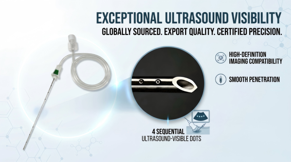
ShineArk Medical's AN-N/AN-M ultrasound-visible nerve block needle series features a 4×4 laser-engraved micro-reflective structure and a 360-degree acoustic reflection design. Understanding the underlying principles helps medical device buyers evaluate these features without confusing a design objective with a guaranteed clinical outcome.
Why Is Needle Visibility Important in Ultrasound-Guided Nerve Blocks?
Ultrasound-guided nerve blocks use real-time imaging to help clinicians assess relevant anatomy and observe needle positioning during a procedure. The ability to distinguish the needle from surrounding structures is an important part of image interpretation.
How Ultrasound Imaging Shows a Needle
An ultrasound probe transmits sound waves into the body and receives echoes from tissue boundaries and other structures. The system processes these echoes to generate an image. The appearance of a needle depends partly on how much sound energy is reflected toward the probe.
When the needle surface is oriented so that reflected sound travels away from the transducer, the needle may appear less conspicuous. Echogenic needle designs aim to modify the interaction between ultrasound waves and the needle surface to support echo return under relevant imaging conditions.
Why Needle Angle Affects Echo Return
A smooth surface can reflect sound directionally, similar to a mirror reflecting light. If the needle is positioned at an unfavorable angle relative to the ultrasound beam, some reflected energy may not return to the probe.
Surface geometry can influence this interaction. However, no surface design should be assumed to eliminate the effects of needle angle, probe orientation, imaging settings, or tissue conditions.
Shaft Visibility Is Not the Same as Tip Identification
A visible section of the needle shaft does not necessarily mean the needle tip is clearly identified. These are separate imaging considerations. Product evaluation should therefore consider both shaft visibility and tip identification under relevant test conditions.
How Does Echogenic Surface Technology Work?
Acoustic Reflection From a Smooth Needle Surface
The amount and direction of reflected ultrasound energy depend on factors including the angle of incidence, surface geometry, material properties, and imaging frequency. A smooth needle surface may produce a strong return under some orientations and a weaker return under others.
How Surface Micro-Structures Affect Echo Return
Echogenic needles incorporate engineered surface features intended to change the way ultrasound waves interact with the needle. Depending on their shape, orientation, spacing, and dimensions, these features can influence the distribution of reflected acoustic energy.
The effect of any particular micro-structure should be supported by product-specific evaluation. It is not appropriate to assume that every patterned surface produces the same result or that a design will provide identical visibility under every imaging condition.
The Relationship Between Needle Geometry and Image Contrast
Needle geometry and surface treatment are only part of the imaging system. Probe frequency, gain, focal position, beam angle, depth, and tissue characteristics also influence the resulting image. For meaningful product comparisons, these factors should be controlled or documented.
How ShineArk's 4×4 Micro-Structure Supports Ultrasound Visibility
ShineArk Medical highlights a 4×4 laser-engraved micro-reflective structure in its AN-N/AN-M ultrasound-visible nerve block needle series. This is a specific product design feature intended to influence the reflection of ultrasound waves.
What Is the 4×4 Laser-Engraved Micro-Reflective Structure?
The 4×4 designation describes the micro-structure arrangement presented by the manufacturer. When discussing this feature, technical content should distinguish the physical pattern from its measured acoustic performance.
Useful supporting materials include close-up product photographs, verified design diagrams, dimensional specifications where available, and test documentation that explains the conditions under which ultrasound visibility was evaluated.
How Is the Structure Distributed Around the Needle Shaft?
The distribution and orientation of reflective features can affect how the surface interacts with incident ultrasound waves. For a specific product, the manufacturer should be consulted for confirmed details about the arrangement and design specifications.
A labeled close-up image of the AN-N/AN-M needle would help readers understand the physical design without relying on broad or unverified performance claims.
What Is the Purpose of the 360-Degree Acoustic Reflection Design?
ShineArk describes its design as a 360-degree acoustic reflection feature. The intended concept is to distribute reflective surface features around the needle circumference to support ultrasound echo return across relevant needle orientations.
The phrase "360-degree" should describe the intended design rather than imply guaranteed visibility at every insertion angle. The actual result should be evaluated under documented imaging conditions.
What Factors Affect Echogenic Needle Visibility?
1. Insertion Angle and Needle Orientation
The relationship between the needle and the ultrasound beam influences how reflected energy returns to the transducer. Visibility may vary when the insertion angle changes, so angle-dependent evaluation can provide useful information about a needle design.
2. Ultrasound Frequency and Probe Position
Probe frequency and position affect image resolution and the way the needle appears. Comparisons should use appropriate imaging settings and document the equipment and probe configuration.
3. Needle Gauge, Surface Pattern, and Tip Geometry
Needle dimensions, surface patterns, and tip configuration may influence the image. Buyers should review the actual model specifications and avoid assuming that results from one size automatically apply to every model in a product family.
4. Tissue Depth and Imaging Settings
Image quality is affected by tissue characteristics, target depth, gain, focus, and other system settings. Results observed in a phantom or demonstration image may not reproduce identically in every clinical setting.
5. Operator Technique and Real-Time Needle Tracking
Probe movement, needle orientation, and image interpretation remain important. Echogenic surface technology is a design aid, not a substitute for appropriate training, procedural technique, or clinical judgment.
How Can Echogenic Needle Visibility Be Evaluated?
Ultrasound Phantom Testing
An ultrasound phantom can provide a controlled environment for comparing needle images. The test setup should document the phantom material, needle model, probe, imaging settings, and insertion angles.
Image Contrast and Needle Tip Identification
Evaluation may consider the visibility of the needle shaft, the ability to distinguish the tip, and the consistency of image interpretation. The method and criteria should be defined before testing.
Comparison Across Different Insertion Angles
Angle-dependent testing can help characterize how the needle appears under different orientations. Where an echogenic needle is compared with a conventional needle, both should be evaluated using comparable conditions and clearly identified test samples.
Repeatability and Test Documentation
Repeat measurements, representative images, test conditions, and documented analysis help buyers understand the basis of a performance claim. Do not infer clinical outcomes from a visual comparison alone.
Frequently Asked Questions
How do echogenic needles improve ultrasound visibility?
They incorporate surface features intended to influence ultrasound reflection and increase the return of echoes to the transducer under suitable imaging conditions.
Why can a needle become difficult to see at steep angles?
The needle surface may reflect ultrasound energy away from the probe. The resulting image also depends on probe position, surface geometry, and imaging settings.
What is the purpose of a 360-degree echogenic design?
It describes a circumferential design approach intended to support ultrasound reflection across relevant needle orientations. It does not guarantee identical visibility at every angle.
Does echogenicity guarantee clear needle tip visualization?
No. Echogenicity is a design characteristic. Tip identification still depends on needle geometry, imaging conditions, and operator technique.
How can buyers compare ultrasound visibility between needle models?
Ask for product-specific images, evaluation methods, test conditions, model specifications, and supporting documentation. Comparisons are most useful when the conditions are clearly described and reasonably consistent.
Explore ShineArk's Ultrasound-Visible Nerve Block Needle Design
ShineArk Medical's AN-N/AN-M series features a 4×4 laser-engraved micro-reflective structure and a 360-degree acoustic reflection design. Buyers can review the available product information and request clarification about model specifications and technical documentation.
Explore AN-N/AN-M Needle Technology
For product evaluation, OEM/ODM discussions, or bulk sourcing, contact ShineArk Medical with your required specifications, intended market, and documentation needs.